AMT / SiO2 Catalyst Characterization Results
- Details
- Category: Tungsten Information
- Published on Wednesday, 24 February 2016 18:27
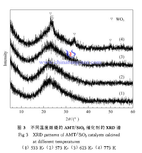
XRD patterns of AMT / SiO2 catalyst calcined at different temperatures can be seen in Figure 3. When the firing temperature is or more than 623K, there is only one large peak dispersion in the XRD spectrum of the sample that SiO2 diffraction peaks without shaped structure of carrier, indicating that particles of tungsten species on the catalyst surface are small or dispersed in a carrier surface in amorphous form. When the firing temperature is 773K, WO3 diffraction peaks appeared in the spectrum of the sample corresponding to the crystal phase, indicating that a high firing temperature treatment can make the AMT catalyst decomposed into WO3.
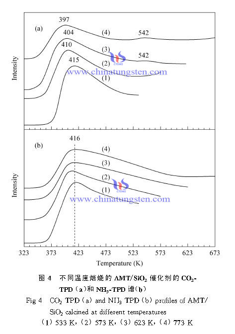
CO2-TPD spectrum of AMT / SiO2 catalyst calcined at different temperatures can be seen in Figure4 (a). The figure shows that a CO2 desorption peak occurs in the vicinity of 410K in the spectra of all sample, this peak slightly moving towards low temperature with increasing calcination temperature, and the peak intensity decreased. It shows that all the samples have a certain amount of presence of a weak base center, and with increasing calcination temperature, the strength and number of weak base centers on the surface of the catalyst are gradually reduced. In addition, there is a small desorption peaks in the samples which the baking temperature is 623 and 773K at about 542K, indicating a small amount of presence of moderate intensity base center in the sample.
Figure 4 (a) is NH3-TPD spectra of the sample. It can be seen in all the samples that NH3 desorption peaks appear about 416K. With the increasing firing temperature, the peak intensity decreased slightly, but the desorption peak temperature substantially unchanged. Which indicates the presence of weak acid center on the catalyst surface calcined at different temperatures, and the amount of weak acid center decreased when calcination temperature increases.
| AMT Supplier: Chinatungsten Online www.ammonium-metatungstate.com | Tel.: 86 592 5129696; Fax: 86 592 5129797;Email:sales@chinatungsten.com |
| Tungsten News&Tungsten Prices, 3G Version: http://3g.chinatungsten.com | Molybdenum News & Molybdenum Price: http://news.molybdenum.com.cn |

 sales@chinatungsten.com